 Image via Wikipedia
Aortic Dissection
Aortic dissection is a condition that is accompanied by severe chest pain. It requires immediate medical attention and usually requires surgery. In the initial stages of aortic dissection, the inner layer of the aorta develops a tear which allows blood to escape and pool at the outer aortic wall. If not treated quickly, the outer wall can rupture, resulting in rapid blood loss. Lack of immediate medical intervention can also cause the tear to spread and block a coronary artery, which leads to myocardial infarction.
Older people are at greater risk for aortic dissection, particularly if they have a history of hypertension, but younger people can develop this condition as well if they have specific inherited defects. One such inherited defect is Marfan's syndrome, which causes a weakening of the aortic wall that can lead to aortic dissection.
Cardiac Arrhythmia
Irregular beating of the heart, also called "arrhythmia," can show up in various ways. Patients may suffer from brief loss of consciousness ("syncope"), experience sensations of irregular or rapid heartbeats ("heart palpitations"), or feel light headed. A careful monitoring of the pulse or a routine ECG exam can detect many such forms of arrhythmias.
In another form of arrhythmia—atrioventricular block—the atrial contraction is not always followed by a ventricular contraction. This causes a slow heartbeat. "Atrioventricular block" can be caused by injury to the pacemaker fibers caused by myocardial infarction or viral diseases.
An abnormally long interval between the Q and the T wave (see the section on ECG) called "long QT syndrome," is another common type of arrhythmia and can occur from a variety of both inherited and non-inherited problems.
An abnormally short interval between heartbeats can be caused by premature contraction of the atrium due to an irregular spread of electrical activity of the ventricle.
Congenital Heart Diseases
When congenital heart disease shows up in the form of a malformed heart at birth, surgical repair is often possible to correct immediate problems.
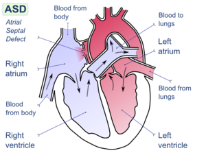
Sometimes, however, congenital heart disease is not detected until adulthood. The most common such problem is an atrial septal defect ("ASD"). In ASD, the septum between the right and left atria has an opening. Typically, because the left heart circulation has higher pressure, blood is shunted from the left to the right atrium, resulting in increased pulmonary circulation and enlargement of the right heart. Symptoms of ASD may include breathlessness and arrhythmia. ASD can be diagnosed by echocardiography and X-ray, and sometimes by ECG.
Other more serious congenital heart diseases include various conditions that lead to cyanosis, in which a substantial mixing of oxygenated and deoxygenated blood occurs. An example of this is "tetralogy of Fallot," which is characterized by an obstruction of the pulmonary outflow combined with a ventricular septal defect, which together allow the mixing of blood between the left and right ventricles.
Myocardial Infarction (MI)
Myocardial infarction (MI), commonly known as a "heart attack," occurs when a patch of heart muscle dies because of a sudden blockage of a coronary artery (coronary thrombosis). It is usually accompanied by crushing chest pain, and often by some evidence of heart malfunction.
Blocking of the coronary arteries is most often due to a condition called "atherosclerosis," which is a buildup of fatty substances in the bloodstream along the inner layer of the arteries over time that restricts blood flow to the heart. These substances can also create an abnormal mass of platelets which become blood clots.
The resulting scar tissue from the dead muscle with MI alters the pattern of electrical activity of the heart. These changes in electrical patterns show up clearly in an electrocardiogram (ECG) exam, making it the most important tool available for diagnosing MI.
Cardiomyopathies
Dilated Cardiomyopathy
This is the most common of the diseases of the heart muscle. In dilated cardiomyopathy, the contraction of the heart muscle is weakened and the left ventricle becomes dilated as a result, leading to reduced blood output.
Dilated cardiomyopathy may develop from many different causes. For example, weakening of the heart muscle can be caused by coronary artery obstruction, which leads to loss of muscle tissue and scarring. Alternatively, infections by agents such as Lyme disease can inflame the heart muscles and cause them to lose strength. Chemicals such as alcohol or cocaine can also lead to this condition.
Dilated cardiomyopathy can be detected most effectively by using an echocardiogram exam to measure ventricular volume, thickness, and function. However, X-ray or ECG exams can also help with detection.
Hypertrophic Cardiomyopathy
In this disease the muscle tissue in the septum that separates the two ventricles become abnormally thickened. The end result is that the ventricular volume is drastically reduced and a smaller amount of blood is ejected from the ventricle at a much higher speed. The speed of ejection and the narrowness of the path of ejection inside the ventricle cause a murmur that can be heard during systole. In addition, the reduced volume of the chamber can interfere with the normal workings of the mitral valve, and mitral regurgitation can occur.
Hypertrophic cardiomyopathy can be caused by hypertension or aortic stenosis, particularly in older people. It may also occur in younger people as a genetically inherited trait known as "familial hypertrophic cardiomyopathy." This disease can often be detected as a heart murmur, but the best diagnostic test is echocardiography, which allows for the measurements of abnormal anatomy and blood flow. The first symptoms of this disease are angina, syncope, ventricular failure, or sudden death.
Valvular Diseases
Aortic Regurgitation
In aortic regurgitation, the aortic valves do not close properly and they allow blood to flow back into the left ventricle. This condition can be caused by diseases of the aortic valves themselves, such as rheumatic heart disease, or by problems with the support structures above the valve, such as aortic aneurysm. The result is a reduction in pumping efficiency, which can lead to dilation of the left ventricle and breathlessness.
Regurgitation can be detected as a blowing murmur (sort of a harsh, whooshing sound), and the timing of the murmur can determine whether it is associated with mitral or aortic valves (unless the valve problems are in the right heart). Echocardiography, particularly the Doppler version showing blood flow, is a very effective technique for diagnosing this condition.
Aortic Stenosis
This obstruction of the outflow from the left ventricle can occur as a defect of the aortic valve itself, or be caused by an obstruction above or below the valve. Obstructed outflow increases the left ventricular pressure and also leads to hypertrophic cardiomyopathy. When valvular aortic stenosis occurs in childhood it is most often congenital, as is most aortic stenosis that occurs above and below the aortic valve. When aortic stenosis occurs in old age it is usually caused by hardening of the valves.
This condition can be detected as murmurs when there is an obstruction of the left ventricular outflow. Usual symptoms for patients suffering from aortic stenosis are angina, breathlessness, and brief loss of consciousness (syncope).
Mitral Regurgitation
Much like aortic regurgitation, mitral regurgitation can be caused by diseases directly affecting the mitral valves. It can also be caused indirectly by other conditions such as hypertrophic cardiomyopathy.
Floppy valve, also known as "mitral valve prolapse," can be caused by diseases of the connective tissue such as Marfan syndrome. In such cases, the floppy valve can produce clicking sounds during systole as the valves balloon into the atrium. The actual regurgitation sounds are blowing murmurs during systole (the pumping phase).
Other causes of mitral regurgitation include damage to the muscles attached to the mitral valves, rheumatic disease, and calcification of the opening between the left atrium and left ventricle. Apart from the characteristic sounds, mitral regurgitation can be easily detected by Doppler echocardiography.
Mitral Stenosis
The main cause of mitral stenosis in adults is rheumatic heart disease. In these cases, an acute childhood rheumatic fever causes the mitral valves to become calcified and to thicken, which leads to a progressively narrower opening into the left ventricle.
Rheumatic fever is now rare in developed countries, so most occurrences of this disease are in elderly people who suffered rheumatic fever when young. In developing countries, rheumatic fever is still a problem, and the resulting mitral stenosis can cause problems in young people during conditions of circulatory stress such as pregnancy or extreme physical exertion.
Mitral stenosis can be detected by listening to the heart: the first sound is accentuated, while the second sound is followed by an opening snap of the valves. In addition, a rumbling sound may be heard during diastole (which is when the heart relaxes between beats). Echocardiography may reveal thickened mitral valves.
Inherited Heart Diseases
Many of the diseases or conditions described so far can occur as a result of a genetically inherited trait. This section lists some of these heritable diseases. Many of these diseases are not specific to the heart, but stem from a more generalized problem which happens to affect the heart.Ellis-van Creveld Syndrome
This syndrome represents a rare autosomal recessive trait. The fact that it is found more commonly among the Amish people is explained by the founder effect, which occurs when a gene pool is restricted because the population is relatively isolated and has descended from a few individuals.
Ellis-van Creveld syndrome is characterized by atrial septal defects (see congenital heart defects) which in severe cases result in the affected individual having a single combined atrium. Other manifestations of this disease include extra digits and dwarfism.
Familial Hypertrophic Cardiomyopathy
Discussed in the previous cardiomyopathies section. More than 40 mutations have been identified that cause the familial form of hypertrophic cardiomyopathy. These are inherited as autosomal (non sex-linked) dominant traits.
Holt-Orams Syndrome
While this autosomal dominant disorder can cause arm abnormalities, it also manifests as an atrial septal defect (ASD) of varying severity. The electrical activity of the heart may also be disturbed due to the ASD.
Marfan Syndrome
This is an autosomal dominant disorder of varying severity. The disease affects the connective tissue and results in the abnormal length of the extremities: legs, arms, fingers, and toes. Affected individuals may also have problems with their eyes. Marfan syndrome can cause aortic aneurysm and aortic dissection as well as mitral regurgitation and aortic regurgitation.
Ward-Romano Syndrome
This is a form of long QT syndrome (see cardiac arrhythmia and ECG ) that is inherited as an autosomal dominant trait. At least three different genes have been identified, mutations in which can lead to this disease.
The long QT interval results from abnormal electrical conduction in the heart and can lead to brief loss of consciousness (syncope) and sudden death.
![Reblog this post [with Zemanta]](http://img.zemanta.com/reblog_e.png?x-id=a4f8a7b8-5acb-4656-a3f9-a0bccf64bd2f)


0 comentários
Postar um comentário